Patterns and the visual system of the fruit fly ‘Drosophila’.
Dr Iris Salecker is program leader in the Division of Molecular Neurobiology at the Medical Research Council National Institute for Medical Research in London (now part of the Francis Crick Institute). Her current team studies the mechanisms underlying visual circuit assembly in Drosophila, with a special interest in axon-target and neuron-glia interactions. In this exclusive interview she discusses her ideas and work, and her collaborative project with artist, Helen Pynor, for the ‘Deconstructing Patterns’ exhibition.

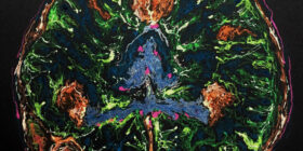
Adult fruit fly optic lobe with Flybow labelled neurons. (Dafni-Hadjieconomou, 2011. Salecker lab MCR National Institute for Medical Research).
Richard Bright: Can we begin by you saying something about your background?
Iris Salecker: I am a senior group leader at the Francis Crick Institute in London, where I am heading the Visual Circuit Assembly Laboratory. I was born in Munich, Germany, and invaluable experiences made while working in laboratories in Germany, France and USA, enabled me to set up my first independent research group here in London at the MRC National Institute for Medical Research, one of the founder institutes of the Francis Crick Institute, more than 17 years ago.
RB: What is the focus of your work?
IS: My team and I are interested in the fundamental question how neural circuits assemble during development to ultimately form a functional brain. We are particularly drawn to the part of the brain dedicated to vision. To find answers, we focus on the visual system of the fruit fly Drosophila melanogaster. This tiny model organism is very powerful to identify the genes that control how nerve cells and another cell type, called glia, are born, acquire their identity and step by step come together as a network. This of course is an enormous task because of the complexity of the nervous system. Therefore, we seek to answer more specific questions, for instance how can nerve cells find their synaptic partners within layers and columns or how do glial cells acquire their remarkable branched morphologies. Because nerve and glial cells face similar tasks in all brains including our own, and many genes between flies and humans are conserved, our findings contribute to increasing our knowledge how the brain develops in general.

Adult fruit fly optic lobe with Flybow labelled neurons. (Dafni-Hadjieconomou, 2011. Salecker lab MCR National Institute for Medical Research.
RB: Can you say something about your Deconstructing Patterns collaboration with Helen Pynor and what it involves?
IS: Bryony Benge-Abbott, the curator of this exhibition, had the wonderful idea to pair scientists at the Francis Crick Institute and artists to explore the theme of patterns. She had been in contact with my PhD student Emma Powell from whom she learned about our research. Deciphering how patterns emerge during development lies at the heart of our work. The regular organization of the fly eye and underlying visual system is simply stunning, and therefore very well suited to convey the concept of patterns. Pairing a lab working on vision with a visual artist therefore seemed a perfect match. To enable us to immerse ourselves into each other’s world, Helen set up her desk and at some point a temporary studio in our lab. Conversely, Helen invited us into her studio in the south of London to tell us about her emerging ideas. Shadowing Emma, she could gain hands-on experience with flies, and brain sample preparations for confocal imaging, and learn about the materials we use. We spent many hours talking about our research over the past months. We undertook experiments together, resulting in the movie clips illustrating the development of the fruit fly, and became also part of the art work itself, resulting in the film capturing my hand gestures to tell the story how the visual system develops. All this has been a truly inspiring and memorable experience.
RB: What questions do you want to address in this collaboration that could not be addressed before?
IS: From the science perspective, we hope to bring across the idea, that developmental biology is a fascinating research area addressing the most fundamental questions about our bodies in general, and the brain, in particular. We also would like to explain how we answer these questions using different types of microscopy in conjunction with genetic and molecular biology techniques. In communicating our findings using our normal ways, we focus on our findings in a factual setting. However, every day we acquire microscopic images, which not only help us to answer specific questions but also are simply beautiful and full of mystery. It is this sense of beauty, wonder and fun, which we hoped to convey through this collaboration. Science is often perceived as not being accessible, and yet, it continues to shape every aspect of our daily lives. Through films, images and voice recordings, the exhibition also seeks to bring across the people behind the science. Everybody has some curiosity for nature in him or herself, why not extend this to the microscopic world? So if visitors of this exhibition get intrigued by this partnership of art and science and science feels a bit more approachable to them, it would be a sign that this unique experiment was successful.

Adult fruit fly optic lobe with Flybow labelled neurons. (Dafni-Hadjieconomou, 2011. Salecker lab MCR National Institute for Medical Research).
RB: What have you personally learnt from working in this collaboration and has this approach thrown up any surprises for you in regards to your previously held beliefs or intuitions?
IS: During this collaboration, Helen and I realized how much our work converged on essentially the same structural problem to convey changes in space and time. Insights gained through our discussions fed into a schematic drawing I was preparing for a recent manuscript, and sharpened interpretations of our findings in this study. Filming parts of the life cycle of our favorite model organism also allowed us for the first time to see the entire sequence of incredible changes that fruit flies undergo during metamorphosis, something only visible to us in snapshots until then. Personally, participating in Helen’s video, I learned about myself, how natural it is for me to use my hands for explaining our research as words never seem to be sufficient to convey ideas. I also began to immerse myself in books exploring the dialogue between art and brain science.
RB: In many ways our human brains interpret information through pattern recognition and re-arranging pattern, which is an evolving dynamic process. What importance does pattern play in your work?
----------------------------------------------------
The rest of this article is reserved for members only. If you have a subscription, please sign in here. Otherwise, why not Subscribe today?
Get the Full Experience
Read the rest of this article, and view all articles in full from just £10 for 3 months.
No comments yet.